In the following interview, the head of the Radiodiagnostic Department, Kamil Sukovský, MD, describes the new device.
How does the new device differ from the original one?
The previous device had 16 rows of detectors, the new one has 128, which significantly improves the spatial resolution and also the speed: the entire chest and abdomen can be covered in a few seconds.
Thanks to state-of-the-art technology, the device can significantly reduce the radiation dose to which the patient is exposed by up to 60%. This is particularly important for young children. At the same time, due to its high speed, excellent spatial resolution and the latest innovative post-processing, the device also helps to protect patients from the possible negative effects of intravenously administered contrast agents, which burden the kidneys. However, we often need this substance: it allows us to distinguish various pathologies in organs and tissues in a wide range of examination procedures. The new device can use a very sophisticated algorithm to monitor the rise in the level of contrast substance in the blood and precisely adjust the scanning of the relevant part of the body according to the rise, so that the amount of contrast that enters the patient's body during the examination is essentially minimal.
What range of patients can be examined on the device?
The maximum gentleness of this new device to the human body not only allows the examination of children or the examination of patients with reduced kidney function, but also, for example, facilitates repeat examinations in patients with, for example, cancer or developing lung diseases, after transplants or after complex surgical procedures.
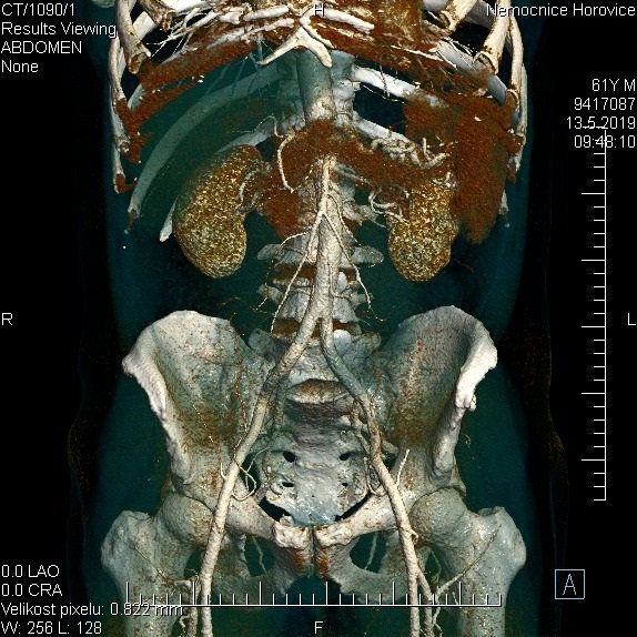
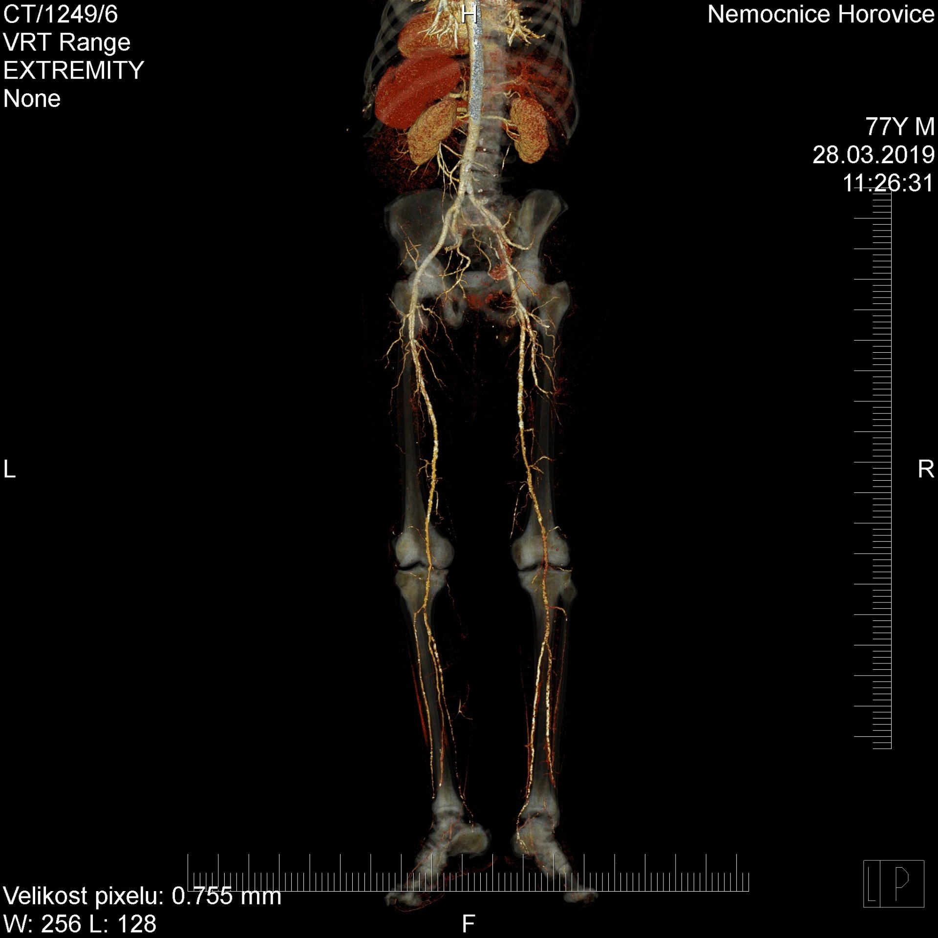
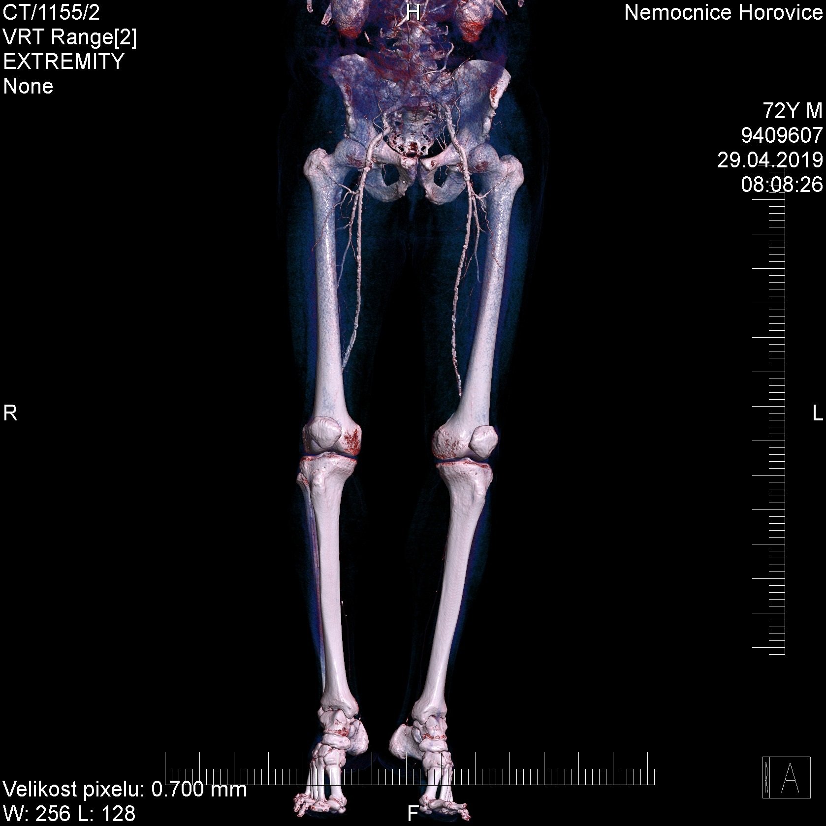
We also perform daily angiographic examinations, particularly of the cerebral arteries, pulmonary arteries and limb arteries, which reveal various blockages and minor narrowing, which can then be used to provide targeted and effective treatment.
The ability of the device to examine the heart is breathtaking. It is amazing how state-of-the-art technology can capture and image even such a troubled organ with such high quality. It uses synchronisation with the ECG to do this and uses high speed and fine resolution. It even displays the individual heart arteries, even in a quality that allows us to quantify any narrowing and plan targeted treatment.
Similarly, the examination of the colon, colonography or so-called virtual coloscopy is very interesting and beneficial, which was also performed at our CT department before, but the new device makes this examination easier and the patient is much better protected from the influence of radiation and the administered contrast agent.
The ability to suppress otherwise very burdensome artefacts from metal joint replacements or other metal bodies is also very welcome.
What other examinations can be performed on the device?
In addition to various examinations, we also perform interventional diagnostic and therapeutic procedures - spraying of nerve roots on the lumbar spine, sampling of pathological deposits, drainage of pathological collections, etc. The new device is a great help in this respect as well: Somatom is equipped with CT fluoroscopy, i.e. the ability to monitor the penetration of the needle or drain into the patient's body on a monitor in real time directly in the examination room and thus precisely target small structures.
The device is also equipped with state-of-the-art post-processing software. This allows, among other things, automatic detection of lung lesions with comparison of their size against the previous CT scan, which remains stored in the device's extensive memory. It can also distinguish certain structures on its own and, for example, directly numbers the vertebrae and ribs, which helps in later orientation. However, it always has all such automatic operations approved or corrected by a live radiologist.
What kind of reconstructions is the software capable of producing?
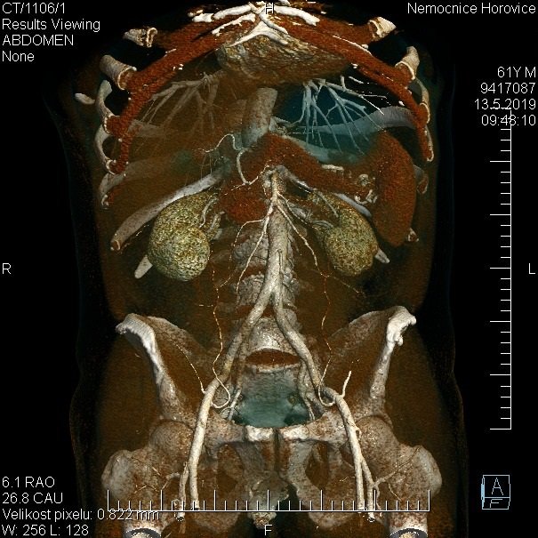
Often the radiologist is quite content with 2D CT slices of the area under examination. But the software's ability to create 3D reconstructions is astonishing. We can position and rotate organs processed with very fine texture on the monitor as needed, and adjust them to either make the pathology even clearer (e.g., the position of fragments of complicated fractures) or to bring the imaged organ or area as close to reality as possible graphically for the clinician or surgeon.
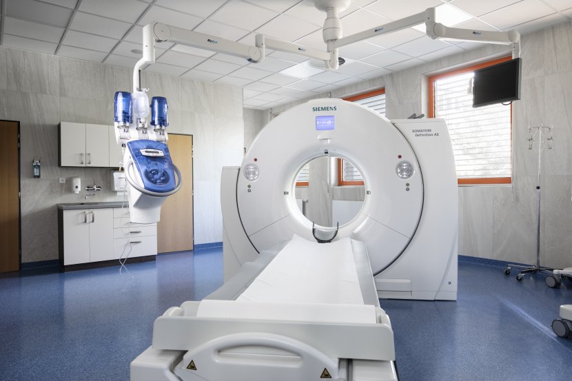
The Siemens Somatom Definition AS+ fundamentally improves patient comfort and safety during examination and diagnosis. The speed of the device and the friendly user environment also means that more patients are examined per unit time than with older CT scans. The resulting image documentation that the radiologist works with is of higher quality and the scan viewer is more ergonomic, so the clinician's work is also accelerated and the resulting benefit to further patient treatment is improved. This means further expanding the ways in which patients can be examined and treated and, for patients, reducing appointment times.


