What is prenatal diagnosis and what is its goal?
Prenatal diagnostics is a set of investigative methods used to diagnose congenital anomalies of the fetus in utero, which allow secondary prevention of congenital malformations. The aim of prenatal diagnosis is to determine the possible presence of fetal congenital malformations during pregnancy and to provide parents with a reasonable indication for treatment of the fetus or termination of pregnancy for severe pathologies based on the information obtained. Prenatal diagnosis involves interdisciplinary collaboration between obstetricians, geneticists, biochemists, neonatologists, cardiologists, radiologists and other specialists and is part of fetal medicine.
What testing methods are used?
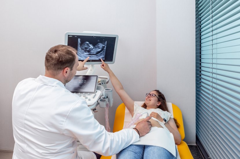
In prenatal diagnosis, we use both non-invasive and minimally invasive methods to obtain information about embryo and fetal development. Non-invasive methods include ultrasound examination, which has an irreplaceable position, and non-invasive prenatal testing (NIPT) from the fetal side. Minimally invasive methods include chorionic villus biopsy (CVS), amniocentesis (AMC), or the less and less used cordocentesis (fetal blood sampling from the umbilical vein).
What ultrasound examinations should every pregnant woman undergo?
Pregnancy care begins with a comprehensive prenatal examination that every pregnant woman undergoes early in her pregnancy. The gynaecologist should recommend screening for the most common fetal VVV and provide the woman with all the information about its methods. Currently in I. Trimester of pregnancy prevails combined screening, which determines the risk of possible occurrence of Down syndrome, Trisomy 13 and 18 in the fetus in the range of 11+0 - 13+6 weeks of pregnancy. The detection rate of screening for Down syndrome is 94%. I definitely recommend screening for the risk of developing pre-eclampsia. This is a disease that appears after the 20th week of pregnancy and can negatively affect the health of the mother and fetus. It is characterised by conditions associated with high blood pressure and associated pathological complications. The test to determine the risk of pre-eclampsia can be carried out in parallel with screening in the first trimester of pregnancy, which has a detection rate of 90%. In the case of increased risk of the disease, preventive therapy is recommended for pregnant women, which significantly reduces the risk of developing and severity of the disease. Ultrasound screening examinations at 18-22 and 30-32 weeks of pregnancy are equally important. For my part, ultrasound screening at 36-37 weeks can certainly be recommended to rule out fetal growth restriction.
Can congenital developmental defects occur at any time during pregnancy or are they only a risk at the beginning?
Congenital developmental defects are pathological deviations from the normal prenatal development of the human individual. Genetic factors, environmental factors, and a combination of both groups are involved in the development of VD. Symptoms of VVV can manifest in varying degrees of severity in both the prenatal and postnatal periods. They represent a relatively wide range of manifestations. Some VVVs are mild, others severely affect the psychomotor development of the stigmatised individual or even cause death.
How common is the occurrence of a congenital developmental defect in a fetus and which defect is the most common?
According to available information, the frequency of fetal VD in the population is approximately 3- 5%. Birth defects with a high incidence include Down syndrome, heart defects or urinary tract defects.
Is there any form of prevention? Can fetal development during pregnancy be influenced?
Prevention of VVV is very difficult, but it is based in any case on planned parenthood and ensuring ideal conditions before pregnancy and subsequently during early embryonic development. Vitamin supplementation and a balanced and varied diet with a healthy lifestyle definitely play a role.