Česky
Česky


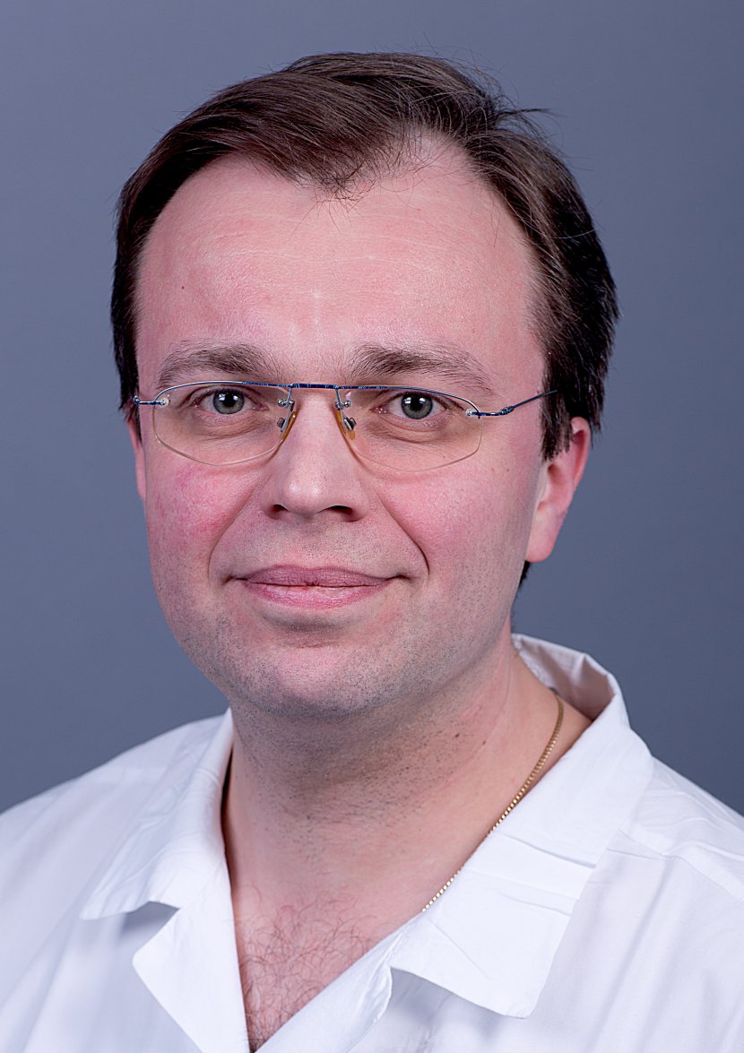
Mr. Chief Medical Officer, you were appointed relatively recently, but you have been working at the hospital in Hořovice for a long time. Could you briefly introduce yourself?
Since I graduated from high school here in Hořovice, I have been more and more eager as a doctor to help people get rid of pain and suffering. In the field I finally chose, the medical profession was combined with one of my hobbies. In fact, my dad led me to a love of the visual arts, and I spent a lot of my childhood looking at encyclopedias and monographs on painting. Radiology as medical imaging became the ideal field for me, combining medicine with my passion for paintings and photographs.
We know that you are a native of Horovice. What is the name of the hospital in your area? Would you recommend it to your friends?
I was born in the hospital in Hořovice. In 1996, after graduation, I joined the hospital and I still work there. I myself was operated on in orthopaedics several years ago, my children have been operated on in surgery, my parents and many of my relatives and acquaintances have been and are being treated here. I know many of the doctors and nurses here and know that they are quality professionals. I can recommend our hospital to everyone with complete confidence as a highly professional facility with quality care.
How is the radiology department in Horovice? What are your priorities in the department?
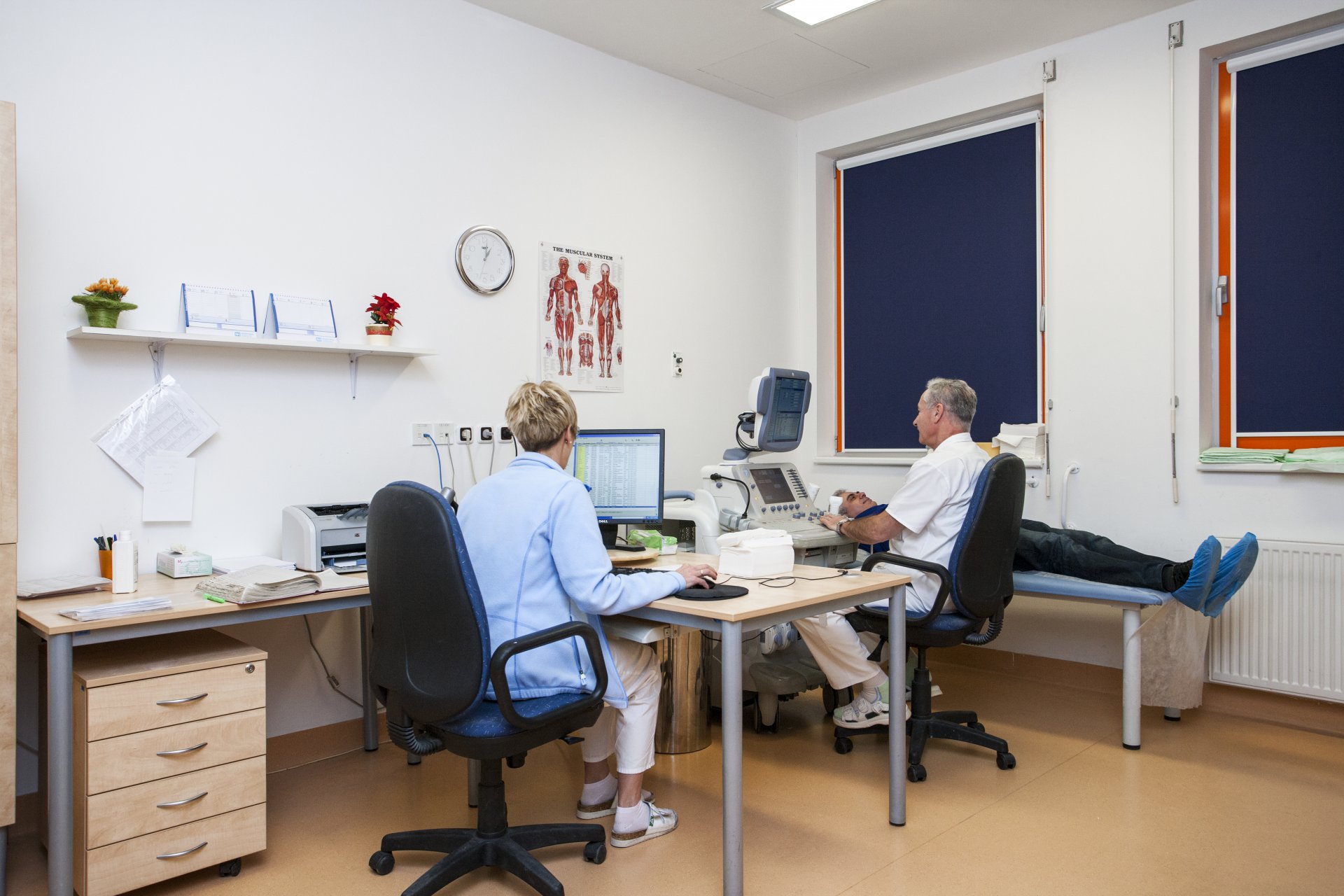
Radiodiagnostics in Hospital Hořovice has imaging methods that are used by virtually all medical disciplines and that help to establish a diagnosis, to assess the course of treatment and the results of therapy. In some cases, we are directly involved in the treatment, for example by interventional methods. Interesting and exciting are the possibilities of X-rays, ultrasound waves or the principle of magnetic resonance to image and evaluate the structures of the human body, tissues and organs and their pathological changes that are hidden deep in the human body.
What innovations does your department offer, or how does it work?
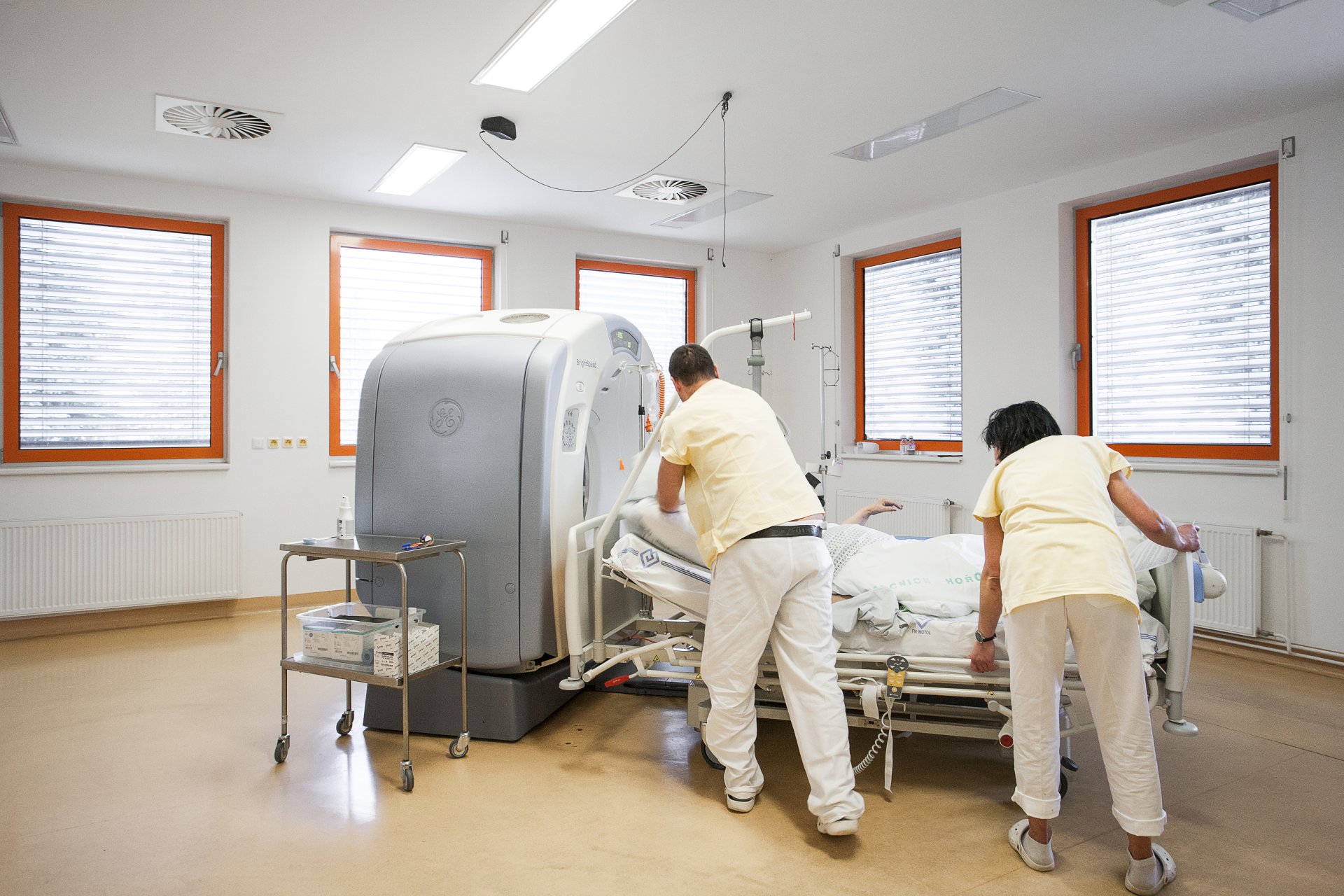
One of the interesting methods is, for example, virtual CT coloscopy. This is a computed tomography (CT) examination of the colon. The examination uses X-rays. The colon is sensitively filled with a small amount of gas before the examination, which causes the walls to distend. An X-ray tube emitting a narrow beam of light is rotated around the patient's body along with a detector, and a computer uses a complex algorithm to create a series of many cross-sections through the patient's body. It then creates a 3D reconstruction that gives us a virtual view of the bowel, allowing us to assess the state of the wall for pathological changes, particularly with regard to cancer prevention. The device then allows the path through the intestine to be followed in further processing, as if the endoscopist were looking through the optics of a colonoscope. We can thus replace conventional colonoscopy to some extent if it is not possible to perform it.